Anatomy Of Musckes Sndctendons / Vintage French Posters Botany Animals Anatomy, old World ... : Each of these muscles is a discrete organ constructed of skeletal muscle tissue, blood vessels, tendons, and nerves.
Anatomy Of Musckes Sndctendons / Vintage French Posters Botany Animals Anatomy, old World ... : Each of these muscles is a discrete organ constructed of skeletal muscle tissue, blood vessels, tendons, and nerves.. Skeletal muscles allow the body to move and maintain posture; • muscle tissues develop from embryonic cells. The muscular system consists of the skeletal muscles and their associated structures. Smooth muscles (involuntary muscles) are usually in sheets or layers, with one layer of muscle behind the other. Skeletal muscle moves bones and other structures.
There are around 650 skeletal muscles within the typical human body. Splenius muscle of head splenius capitis muscle. Learn about the muscles, tendons, bones, and ligaments that comprise the knee joint anatomy. There are over two dozen gorgeous and painstakingly. The anterior and middle scalenes originate from the transverse processes of certain cervical vertebrae and attach to the first rib.
Deep Muscles Of Back Anatomy : 7 Deep Muscles Of Back ... from pulpbits.net Skeletal muscles allow the body to move and maintain posture; Inflammation of this region caused by repetitive stress or trauma may lead to pain and numbness known as carpal tunnel syndrome. Find the best weight lifting exercises that target each muscle or groups of muscles. By contracting, they also aid the venous return of blood to the heart and with age, these components of the musculoskeletal system progressively degenerate, which contributes to frailty and increases the risk of falls and fractures. The tendons of these muscles pass through a small corridor in the wrist known as the carpal tunnel. Muscle tendons are extremely important in reinforcing and stabilizing joints. From anterior to posterior, the tongue has 3 surfaces: The anterior and middle scalenes originate from the transverse processes of certain cervical vertebrae and attach to the first rib.
Muscle movements, types, and names.
See the pictures and anatomy description of knee joint bones, cartilage, ligaments, muscle and tendons with resources for knee problems & injuries. The tendons of these muscles pass through a small corridor in the wrist known as the carpal tunnel. Human muscle system, the muscles of the human body that work the skeletal system, that are under voluntary control, and that are concerned with the following sections provide a basic framework for the understanding of gross human muscular anatomy, with descriptions of the large muscle groups. There are two main muscle groups around the knee: The tongue is a mass of muscle that is almost completely covered by a mucous membrane. Splenius muscle of head splenius capitis muscle. Knee function is determined in large part by the anatomy of the joint. Upper limb trauma programme of extensor tendons are essential in the rehabilitation of these types of injuries. Attached to the bones of the skeletal system are about 700 named muscles that make up roughly half. Convergent muscles contain fibers that have a wide origin, but converge in order to attach to a narrow tendon. The muscles around the knee help to keep the knee stable, well aligned, and moving. Muscle movements, types, and names. Inflammation of this region caused by repetitive stress or trauma may lead to pain and numbness known as carpal tunnel syndrome.
The muscular system consists of the skeletal muscles and their associated structures. The muscular system is responsible for the movement of the human body. Skeletal muscles are attached to bones by tendons and can be as long as 30 cm, although they are usually 2 to 3 cm in length. The primary function of the knee is to hinge at the lower extremity. The smooth muscle tissue that forms organs like the stomach and bladder changes.
Muscles of the Neck and Trunk - Learn Muscles from learnmuscles.com The muscles around the knee help to keep the knee stable, well aligned, and moving. Microscopic anatomy of skeletal muscle. Smooth muscles (involuntary muscles) are usually in sheets or layers, with one layer of muscle behind the other. Muscle movements, types, and names. The smooth muscle tissue that forms organs like the stomach and bladder changes. • muscle tissues develop from embryonic cells. Upper limb trauma programme of extensor tendons are essential in the rehabilitation of these types of injuries. Anatomy of the muscular system.
There's no strict demarcation or dividing line between the tendon and the covering around this muscle but that covering is called is called the epimysium fp my cm and it's really just connective tissue that covers the muscle kind of protects it reduces friction.
Muscle movements, types, and names. How to study muscle anatomy. The three scalene muscles are found forming the floor of the posterior triangle. Anatomy of the short head of the biceps brachii muscle. There's no strict demarcation or dividing line between the tendon and the covering around this muscle but that covering is called is called the epimysium fp my cm and it's really just connective tissue that covers the muscle kind of protects it reduces friction. Muscle mass accounts for a large majority of the carcass weight of domestic animals. Muscle tendons are extremely important in reinforcing and stabilizing joints. The tendons of these muscles pass through a small corridor in the wrist known as the carpal tunnel. Splenius muscle of head splenius capitis muscle. The tip is the highly mobile, pointed anterior portion of the tongue. Muscular contraction is necessary for voluntary and involuntary movement of limbs, stabilization of joints, maintaining luminal diameter (in the case of arteries, bowel, etc), and to produce heat. The primary function of the knee is to hinge at the lower extremity. An interactive tutorial teaching the position, actions, innervation and attachments of the rectus femoris muscle with the aid of anatomical illustrations.
Anatomy of a muscle cell. Learn about the muscles, tendons, bones, and ligaments that comprise the knee joint anatomy. See the pictures and anatomy description of knee joint bones, cartilage, ligaments, muscle and tendons with resources for knee problems & injuries. The muscles of the torso, examined in the previous chapter, include a few that attach directly into the upper arm and help move the humerus at the shoulder joint. An interactive tutorial teaching the position, actions, innervation and attachments of the rectus femoris muscle with the aid of anatomical illustrations.
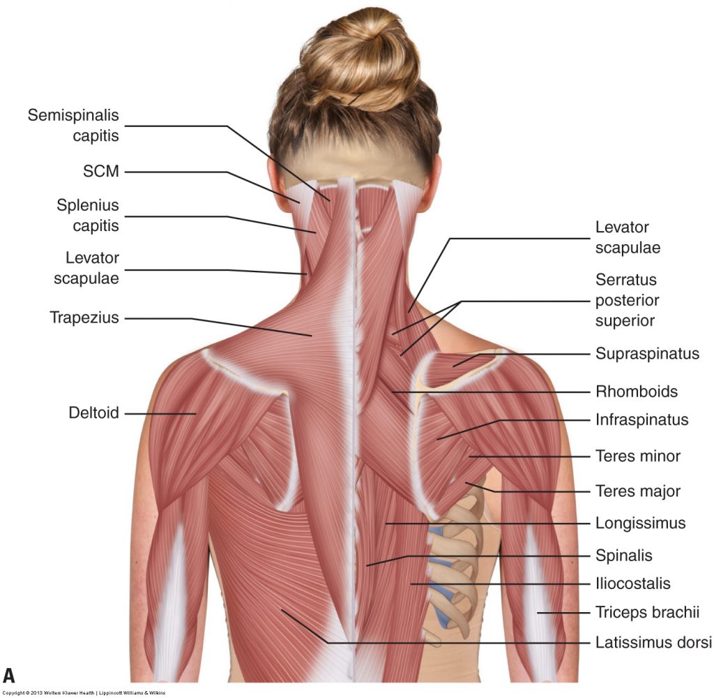
Muscle Chart: Anatomical Muscle Chart - SteroidsLive from www.steroidslive.com You can click the links in the image, or the links below the image to find out more information on any muscle group. Find the best weight lifting exercises that target each muscle or groups of muscles. Learn about human anatomy muscles with free interactive flashcards. Topographically, the muscles in this group are classed along with the lateral torso wall and upper extremity, which is due to their location as well as their genetic development based on their embryological origin. There are over two dozen gorgeous and painstakingly. In this section, learn more about the anatomy of the muscles of the neck. Muscle movements, types, and names. Splenius muscle of head splenius capitis muscle.
Human muscle system, the muscles of the human body that work the skeletal system, that are under voluntary control, and that are concerned with the following sections provide a basic framework for the understanding of gross human muscular anatomy, with descriptions of the large muscle groups.
Knee function is determined in large part by the anatomy of the joint. Microscopic anatomy of skeletal muscle. Muscle tendons are extremely important in reinforcing and stabilizing joints. Related online courses on physioplus. The tendons of these muscles pass through a small corridor in the wrist known as the carpal tunnel. Learn about the muscles, tendons, bones, and ligaments that comprise the knee joint anatomy. Anatomy of the muscular system. An interactive tutorial teaching the position, actions, innervation and attachments of the rectus femoris muscle with the aid of anatomical illustrations. Muscle movements, types, and names. Skeletal muscles are attached to bones by tendons and can be as long as 30 cm, although they are usually 2 to 3 cm in length. Inflammation of this region caused by repetitive stress or trauma may lead to pain and numbness known as carpal tunnel syndrome. Attached to the bones of the skeletal system are about 700 named muscles that make up roughly half. The primary function of the knee is to hinge at the lower extremity.


0 Komentar